ApproachMay vary
Interlaminar, transforaminal, or caudal
The procedure name alone does not establish the approach, target level, or side. Those details come from the current order and responsible clinician.
Understand the procedure, the room, the team, and what deserves confirmation.
LESI stands for lumbar epidural steroid injection. It is an image-guided procedure in which a credentialed clinician places ordered medication into the epidural space of the lower spine. It may be considered when lower-spine nerve irritation is associated with pain or related symptoms; the responsible clinician determines whether it is appropriate.
Medication status: The FDA has not approved injectable corticosteroids for epidural administration. Material risks and patient-specific counseling belong to the responsible clinician and facility consent process. Review the free safety orientation.
General clinical-team orientation. Current orders, qualified-clinician direction, facility policy, and approved product information remain controlling.
Start with the purpose and the basic process.
The procedure name alone does not establish the approach, target level, or side. Those details come from the current order and responsible clinician.
It may be considered for pain that travels from the lower back into the leg or for symptoms linked to nerve irritation. The treating clinician decides whether it is appropriate.
The patient is positioned, the skin is cleaned and numbed, and live imaging guides the epidural needle. Contrast commonly helps confirm location before ordered medication is delivered.
See the usual setting, position, and equipment.
This may be a hospital outpatient department, pain clinic, ambulatory procedure center, or physician office equipped for image-guided procedures.
Supports may be used for comfort and access. Patient needs and the planned approach determine the actual position.
A fluoroscopy C-arm or CT system, compatible table, image monitor, positioning supports, and sterile setup are commonly present. Monitoring equipment depends on the case.
These details can change and should come from current approved sources.
An appropriately qualified clinician performs the injection. Nursing, radiologic technologist, monitoring, recovery, and sedation support depend on the setting and plan.
Exact needle, syringe, skin preparation, drape, and contrast-delivery selections depend on the approach and the facility’s approved source.
Confirm each medication, formulation, concentration, dose, route, allergy, expiration, and sequence from current orders and approved sources. Corticosteroids are not FDA-approved for epidural administration.
A conceptual view of common room elements—not a fixed staffing model, workflow, or depiction of technique.
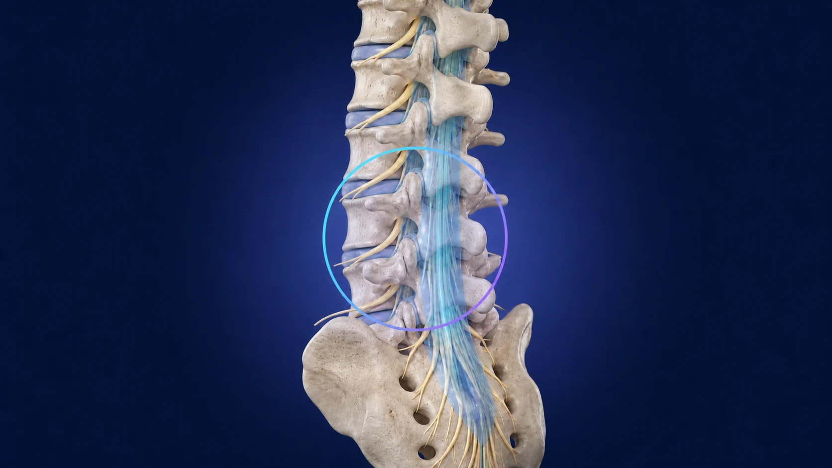
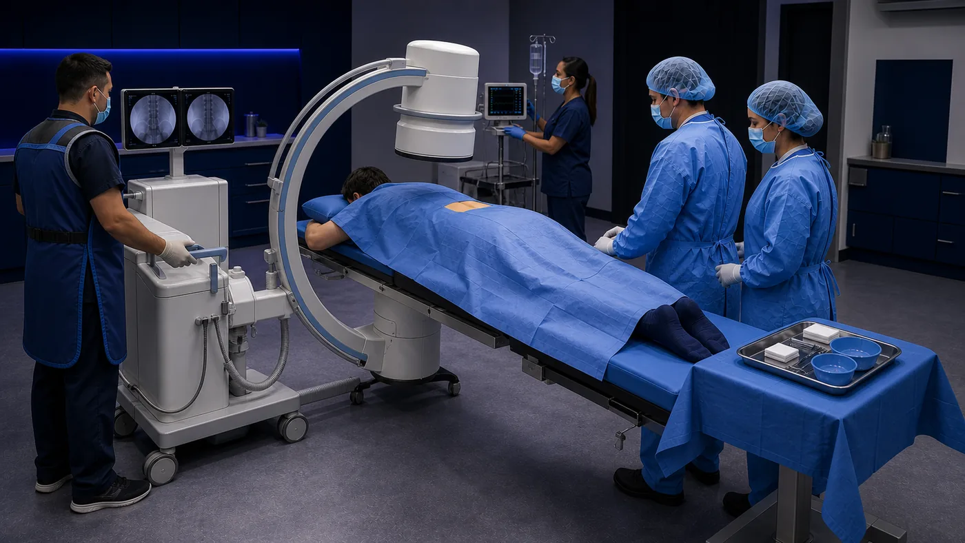
Original AI-generated VSEEE educational illustrations. They provide general anatomy and room orientation and do not establish an exact procedural site, required setup, or technique.
Descriptive orientation—not a protocol, checklist, or set of clinical instructions.
Confirm the lumbar region, indication, intended approach, level and side when specified, current imaging, consent pathway, allergies, medication plan, bleeding review, infection screening, and post-procedure arrangements.
After positioning and sterile preparation, local anesthetic is commonly administered. The qualified clinician advances the needle using the selected imaging method, confirms placement according to the ordered approach and local process, and administers the ordered medication.
Monitoring, neurologic assessment, activity guidance, glucose considerations, transportation, discharge criteria, and instructions for reporting new or worsening symptoms follow the responsible clinician’s and facility’s requirements.
These prompts do not replace the facility checklist, consent, product information, or responsible-clinician direction.
The free briefing explains the procedure. Complete continues with the LESI room, equipment, instruments, supplies, medication-source entries, staff roles, and universal readiness areas in one printable guide.
Use the current source and approved local documents for decisions; this page is a concise orientation.
ACR–ASNR–ASSR–SIR–SNIS Practice Parameter for the Performance of Image-Guided Epidural Steroid Injection, revised 2025.
Patient-facing overview developed by the American College of Radiology and Radiological Society of North America.
The FDA warns of rare but serious neurologic events and states that corticosteroids are not approved for epidural administration.
CDC guidance addresses single-dose containers, one-patient use, aseptic technique, and injection safety for spinal procedures.
DEA guidance describes registration and ordering requirements for entities authorized to handle controlled substances.